Back Of Head Skull Anatomy - Understanding Spine Anatomy Rojeh Melikian M D - It is a complex anatomical structure weighing up to five kilograms that rests on the bony skull and in turn, the neck.
byAdmin•
0
Back Of Head Skull Anatomy - Understanding Spine Anatomy Rojeh Melikian M D - It is a complex anatomical structure weighing up to five kilograms that rests on the bony skull and in turn, the neck.. It is also known as the calvarium. The erector spinae are a group of many muscles that attach along the back of the spine. We will look at the workings of this tiny portion of the skull. In addition to the evident ears, eyes, nose, and mouth, the head supports a variety of other important structures: Usually, headaches in the back of head are the result of stress, muscle tightness, tension, the overuse of medications, and tiredness.
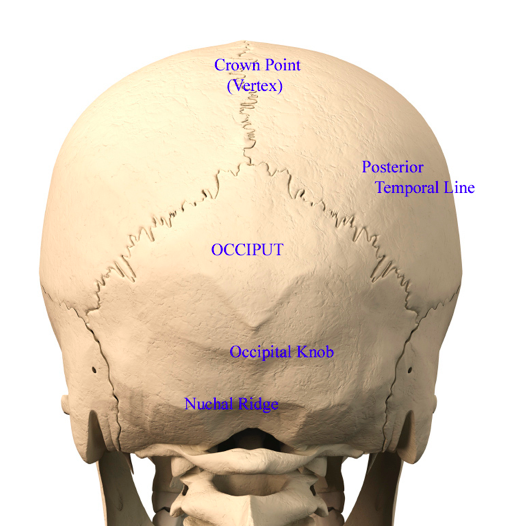
The occipital bone (/ ˌɒkˈsɪpɪtəl /) is a cranial dermal bone and the main bone of the occiput (back and lower part of the skull). It is comprised of many bones, formed by intramembranous ossification, which are joined together by sutures (fibrous joints). Sometimes a pain in the base of your skull can be caused by occipital neuralgia which is a condition that affects the nerves that run from the top of the spinal cord up through the scalp. As one of the 22 skull bones, this bone behind the ear is the connection point for major neck and head muscles, nerves, and tissues. See human skull anatomy stock video clips.
Occipital Neuralgia And Suboccipital Headache C2 Neuralgia Treatments Without Nerve Block Or Surgery Caring Medical Florida from www.caringmedical.com This is the second video about the skull anatomy and the bones of the head. Skeleton peeking through an old truck bed suspiciously keeping an eye on things back view. Herniated disks in the cervical spine (neck) can cause neck pain and tension. The skull is a strong, bony capsule that rests on the neck and encloses the brain. The orbicularis oris is a circular muscle that moves the lips, and the orbicularis oculi is a circular muscle that closes the eye. Finally i explained the skull anatomy in my series about the head anatomy. An area called the occiput. Human anatomy for muscle, reproductive, and skeleton.
Learn about the anatomy of the skull bones and sutures as seen on ct images of the brain.
Knowledge of the anatomy of the vasculature of the head and neck from the thorax to the skull base is critical to the approach to diagnosis and treatment of cerebrovascular disease. Learn about the anatomy of the skull bones and sutures as seen on ct images of the brain. There are many muscles around the neck that help to support the cervical spine and allow you to move your head in different directions. Ct anatomy of skull, axial reconstruction, bone window. Learn about the anatomy of the skull bones and sutures as seen on ct images of the brain. This can cause a type of headache called a cervicogenic headache. See anatomy of the head and neck stock video clips. The erector spinae are a group of many muscles that attach along the back of the spine. Learn about the anatomy of the skull bones and sutures as seen on ct images of the brain. The orbicularis oris is a circular muscle that moves the lips, and the orbicularis oculi is a circular muscle that closes the eye. The nerves of the head and neck include the most vital and important organs of the nervous system — the brain and spinal cord — as well as the organs of the special senses. 12 photos of the bone of back of skull bone back of head bigger on one side, bone back of the head, bone on back of skull, bone spur back of skull, skull bone back of head, bone, bone back of head bigger on one side, bone back of the head, bone on back of skull, bone spur back of. Human anatomy for muscle, reproductive, and skeleton.
What is the pain in back of head at base of skull ? They move the head in every direction, pulling the skull and jaw towards the shoulders, spine, and scapula. Knowledge of the anatomy of the vasculature of the head and neck from the thorax to the skull base is critical to the approach to diagnosis and treatment of cerebrovascular disease. The erector spinae are a group of many muscles that attach along the back of the spine. It might only be due to a minor injury or it can be a secondary symptom of other problems in the body.
Back Of Head Skull Anatomy Dr Barry Eppley Indianapolis Explore Plastic Surgery from exploreplasticsurgery.com Sometimes a pain in the base of your skull can be caused by occipital neuralgia which is a condition that affects the nerves that run from the top of the spinal cord up through the scalp. The skull is the bony skeleton of the head. As one of the 22 skull bones, this bone behind the ear is the connection point for major neck and head muscles, nerves, and tissues. Learn about the anatomy of the skull bones and sutures as seen on ct images of the brain. Working in pairs on the left and right sides of the body, these muscles control the flexion and extension of the head and neck. In medical term, it is called as occipital neuralgia. With the mastoid process location being close to the ear, any infection of the ear or blow to this region of the head may damage this vital bone. The foramen magnum, housing the brainstem, is also a part of the occipital bone.
Working in pairs on the left and right sides of the body, these muscles control the flexion and extension of the head and neck.
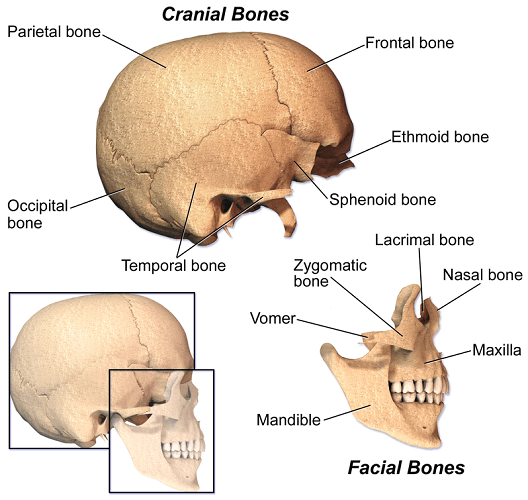
As one of the 22 skull bones, this bone behind the ear is the connection point for major neck and head muscles, nerves, and tissues. One of the more common types of cancerous skull tumor is a chordoma, which is a tumor that can grow from the bones at the base. The occipital bone houses the back part of the brain and is one of seven bones that come together to form the skull. The occipital bone is a bone that covers the back of your head; Knowledge of the anatomy of the vasculature of the head and neck from the thorax to the skull base is critical to the approach to diagnosis and treatment of cerebrovascular disease. The skull is the bony skeleton of the head. The neurocranium (cranial vault) and the viscerocranium (facial skeleton). This can cause a type of headache called a cervicogenic headache. The occipital bone surrounds a large opening known as the foramen magnum. The erector spinae are a group of many muscles that attach along the back of the spine. The trapezius originates from the skull and spine of the upper back and neck. A thorough description is beyond the. The orbicularis oris is a circular muscle that moves the lips, and the orbicularis oculi is a circular muscle that closes the eye.
Dents and irregularities in the shape of your skull are usually simple variations in anatomy. There are many muscles around the neck that help to support the cervical spine and allow you to move your head in different directions. The erector spinae are a group of many muscles that attach along the back of the spine. The pain typically originates and is felt in the back. Nerves of the head and neck.
The Skull The Definitive Guide Biology Dictionary from biologydictionary.net Herniated disks in the cervical spine (neck) can cause neck pain and tension. What is the pain in back of head at base of skull ? You don't give your age or how long you have had this indentation. Headaches in the back of the head can have a number of different causes; See anatomy of the head and neck stock video clips. It consists of two major parts: Nerves of the head and neck. The trapezius originates from the skull and spine of the upper back and neck.
12 photos of the bone of back of skull bone back of head bigger on one side, bone back of the head, bone on back of skull, bone spur back of skull, skull bone back of head, bone, bone back of head bigger on one side, bone back of the head, bone on back of skull, bone spur back of.
The type and location of the pain can play a crucial role in diagnosing the cause of headaches.severe and recurrent headaches always require medical attention from a doctor. If it's recent, did you fall? This is the second video about the skull anatomy and the bones of the head. The muscle has a frontal belly and an occipital (near the occipital bone on the posterior part of the skull) belly. Learn about the anatomy of the skull bones and sutures as seen on ct images of the brain. What is the pain in back of head at base of skull ? The erector spinae are a group of many muscles that attach along the back of the spine. We will look at the workings of this tiny portion of the skull. It is comprised of many bones, formed by intramembranous ossification, which are joined together by sutures (fibrous joints). A thorough description is beyond the. An area called the occiput. The occipitofrontalis muscle moves up the scalp and eyebrows. Everyone has variations in bone structure — just consider how very different people's faces can look.
With the mastoid process location being close to the ear, any infection of the ear or blow to this region of the head may damage this vital bone back of skull anatomy. The type and location of the pain can play a crucial role in diagnosing the cause of headaches.severe and recurrent headaches always require medical attention from a doctor.